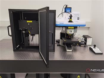
Raman Spectrometer - Confocal Raman Microscope XploRA PLUS-OmegaScope
Contact:
Assist. Zoran Lavrič, M. Pharm., Ph. D.
Access:
Equipment is available at the Faculty of Pharmacy, Aškerčeva 7, 1000 Ljubljana. Access to equippment must be agreed with supervisor of the equipment. Due to delicate nature of the equipment supervisor must be present through whole ageed working time on the equipment.
Intended Purpose:
The integrated system of the confocal Raman microscope and the atomic force microscope allows various methods of microscopic analysis of the sample to be performed. The confocal microscope enables the observation and capture of a visible microscopic image. Laser light sources (532 nm and 785 nm), associated optical elements and Raman backscatter detector enable point measurements of properties on the principle of Raman spectroscopy and photoluminescent spectroscopy in two and three dimensions, which allows microscopic Raman mapping of the surface and volume of the sample surface and cross-sections of bulk free particles, granules, pellets, tablets, capsules, films, laminates, extrudates, etc.) and in a liquid medium (fibers, particles, cells, tissues, etc.).
Description of equipment:

Raman measurements are performed in reflectance mode, where the sample is irradiated with a laser beam through a lens above the sample and then Raman scattered laser beams are collected through the same lens and their spectral properties are determined. The integrated microscope system has a special module for mapping the surface of bulk free particles, which is performed according to a sample - adapted procedure, followed by processing the captured data with appropriate pre - treatment, segmentation and evaluation, which allows classification of particles according to size, different dimensional ratios (Ferret, etc. ) or spectral properties. The Atomic Force Microscope (AFM) module enables the measurement of the topographic, adhesive and mechanical properties of the surface of pharmaceutical samples in various derivatives of the contact mode, tapping and non-contact mode. Sensory microscope measurements can be performed in a thermostated aqueous medium (between 25 ° C to 60 ° C) for the purpose of studying cell surface interactions. The software enables instrument control, creation of procedures for mapping, capture and storage of 2D, 3D and hyperspectral data, data processing and evaluation of optical surface maps, hyperspectral maps and AFM maps (pre-processing, multivariate analysis), 2D and 3D presentation of results and correlation of Raman and AFM surface maps.
Price of equipment use
Use is 29,51 € per hour.
Research Equipment ( co)funded by the Slovenian Research Agency.
Changed: 27. January 2021